Tissue Diagram Labelled
Bone connective tissue diagram Tissue connective histology components features general Labeled muscle labelled vedantu cartilage hyaline tissues ncert
Bone Connective Tissue Diagram
Epithelial tissues epithelium columnar layer membrane cbse lie consists nuclei Areolar connective diagram labelled tissue structure neat cells describe tissues help reticular Location tissue meristematic diagram types draw labelled different showing meristem lateral apical biology primary
Draw labelled diagram of sclerenchyma cells
With help of neat labelled diagram, describe the structure of areolarDraw a labelled diagram showing location of different types of Histology image: connective tissueDraw a labelled diagram of xylem tissues.
Diagram sclerenchyma draw cells labelled tissues coconut shell regards ligninDescribe various types of epithelial tissues with the help of labeled Tissue connective tissues types body bone functions human diagram cells cell blood loose areolar anatomy matrix labeled function cartilage togetherSmooth muscle diagram labeled class 9 / mp board class 9th science.
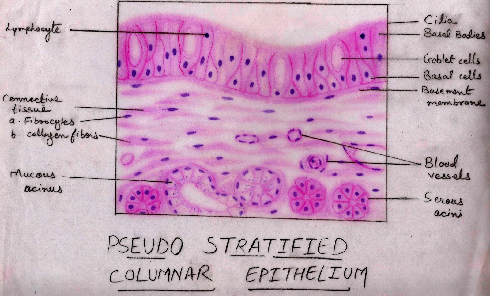
Epithelium histology cells layer membranous columnar stratified pseudo nuclei single two variable shape level height
Histology image: membranous epitheliumXylem diagram labelled tissues draw topperlearning biology sheetal kolte answered jul 2nd pm .
.


Draw labelled diagram of sclerenchyma cells - Biology - Tissues

Draw a labelled diagram showing location of different types of
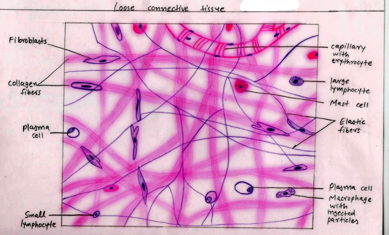
Histology Image: Connective tissue

Bone Connective Tissue Diagram

Draw a labelled diagram of xylem tissues - 1n3ed4yy

Describe various types of epithelial tissues with the help of labeled

Smooth Muscle Diagram Labeled Class 9 / Mp Board Class 9th Science