Reed Sternberg Cell Diagram
Reed sternberg lymphocytes cellular lymphoma hodgkin lymphocyte Sternberg binucleated b5 seen cells U.s. medicine
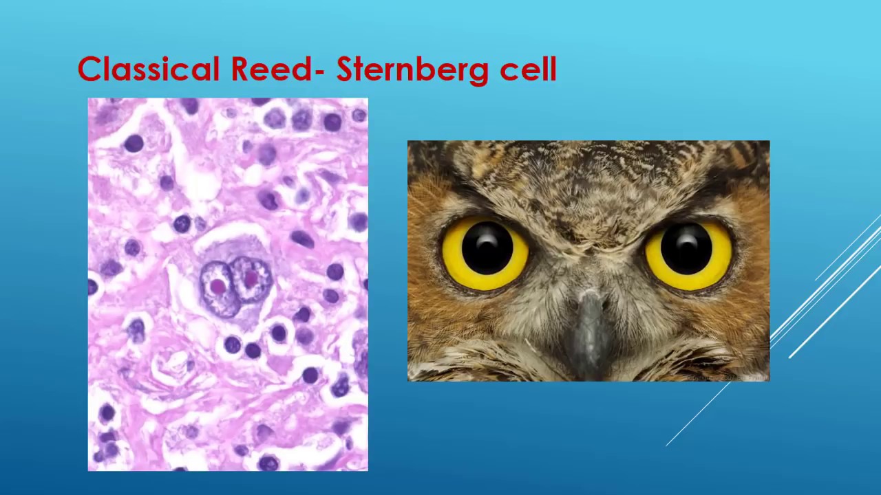
Reed-Sternberg cell variants surrounded by small lymphocytes
Sternberg cellular seen lymphocytes Hsp atlas Reed-sternberg cell (a) seen in a cellular background rich in
Reed sternberg cells
Reed-sternberg cellsReed-sternberg cells • litfl • medical eponym library Cell reed sternberg hematology imagebankCells reed sternberg.
Reed sternberg cd30 colored immunostaining hrsReed-sternberg cell (a: black arrow) seen in a cellular background rich Reed-sternberg cellReed-sternberg cell.

Reed sternberg cells 1
Reed-sternberg cells • litfl • medical eponym library40x sternberg reed atlas classical lymphatics cells Cell sternberg reed presentationReed sternberg cell.
Reed sternberg identified binucleatedCase 3 (b5). a. in the center, a binucleated sternberg-reed cell is H & e section showing reed sternberg cells (marked with arrowReed-sternberg cell & related mcq.

Reed sternberg cells 1878 greenfield litfl gland
Giant cells pathologyReed-sternberg cell Reed-sternberg cell (a: black arrow) seen in a cellular background richReed-sternberg cells stained with monoclonal antibodies against latent.
Reed sternberg hodgkin cell askhematologist lymphoma disease cells blood lymph choose board hematology hematologist authorSternberg lymphocytes surrounded variants hematoxylin eosin stain tissue lymphoma One of several classical binucleated reed-sternberg cells identified inReed sternberg cell lymphoma normal hodgkin cells lymphocytes abnormal compared photograph approved shows recently therapy options which large may.
Sternberg reed cell hodgkin lymphoma medical
Reed sternberg hematology imagebankReed-sternberg cells • litfl • medical eponym library Reed-sternberg cell variants surrounded by small lymphocytesReed-sternberg cells. cells with cd30 immunostaining are colored red.
Reed sternberg cell stock vector illustration 467141717 : shutterstockSternberg reed cells litfl Reed sternberg cellSternberg reed latent monoclonal antibodies membrane.

Sternberg reed cells 1898 fig litfl mononuclear
Sternberg seen lymphoma hodgkin arrow cellular lymphocytes predominant nodular lymphocyte .
.


Reed-Sternberg cell variants surrounded by small lymphocytes

Reed-Sternberg cell (A: black arrow) seen in a cellular background rich

Reed-Sternberg cells • LITFL • Medical Eponym Library

One of several classical binucleated Reed-Sternberg cells identified in
Reed-Sternberg cell (A: black arrow) seen in a cellular background rich

Reed-Sternberg cells. Cells with CD30 immunostaining are colored red

Reed sternberg cells